Digging Tunnels: Cytotoxic T Lymphocytes Mechanisms of Migration
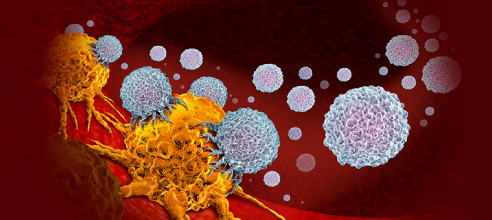
Cytotoxic T lymphocytes (CTLs) are cells that belongs to the adaptive immune system which eliminate tumorigenic or pathogen-infected cells. The process by which CTLs recognize the cognate antigens presented by target cells to eliminate them is called immunosurveillance. As target cells are often in low number in the early stages of disease development [1], for an efficient immune response CTLs must efficiently navigate and search within the tissues. It has been described that migration of naive T cells within lymph nodes follows a Brownian or even subdiffusive dynamics [2], but switching between fast and slow motility modes have also been observed [3]. Peripheral tissues, in which activated T cells must find their targets, are characterized by a dense extracellular matrix (ECM) where a faster migration is advantageous to scan a larger tissue more efficiently. The ECM mainly consist of collagens, some of which have inhibitory effects on the function of different immune cells [4]. Therefore, when in the vicinity of tumors, the collagen network becomes stiff, dense and linearized the migration of cancerous cells is facilitated and the proliferation of CTLs is impaired [5], thus making the ECM an important player in cancer metastasis, prognosis and immunosurveillance escape.
Understanding the migration and interactions of immune cells within collagen networks is crucial to unravel the underlying details of immune response and design of effective treatment strategies.
Trajectories of CTLs in 3D collagen matrices. Sadjadi Z, et al. Migration of Cytotoxic T Lymphocytes in 3D Collagen Matrices. Biophys J. 2020 Dec 1;119(11):2141-2152. doi: 10.1016/j.bpj.2020.10.020
In a recent article published in Biophysical Journal [6], Zeinab Sadjadi, et al used collagen hydrogels to compare the migration patterns of human CTLs in aligned and nonaligned collagen fibers microenvironments, which resembles tumor cells and normal tissues, respectively. Migration of CTLs can be broadly categorized into slow, fast and mixed subgroups plus a persistent random walk model with two different motility states: slow and fast. Authors investigate the migration patterns of primary human CTLs in a 3D microenvironment by embedding cells into collagen matrices and visualizing their movements using light-sheet microscopy. They defined three different types of CTL trajectories: 1) slow cells that perform a subdiffusive motion with velocities that always remain below a threshold value, 2) a faster group with velocities above a threshold value, and 3) a group with velocities that switch between fast and slow modes. These results allow researchers to define three classes of trajectory types: fast, slow and mixed. When increasing collagen density, they observed a moderate increase of the average persistence and a moderate decrease of the average velocity, being faster T cells more persistent than the slow ones in a density-independent manner.
The mean-square displacement (MSD) of different types of motion are clearly distinguishable, and the differences in the level of MSD curves reflect the differences in the average velocity of CTLs in various migration types. The MSD of the mixed type coincides with the total MSD in nearly all cases, showing that the mean velocity of all T cells is nearly the same as the mean velocity of the mixed type when the resident times in the substrates of the mixed type are taken into account.
MSD of different motility types. Blue dots represent slow motility, green triangles fast motility and yellow squares the mixed one. The solid line represents the MSD of all T cells. Sadjadi Z, et al. Migration of Cytotoxic T Lymphocytes in 3D Collagen Matrices. Biophys J. 2020 Dec 1;119(11):2141-2152. doi: 10.1016/j.bpj.2020.10.020
They also found that the sojourn times (the time that a T cell remain in one state before they switch) follow an exponential decay, indicating that the transition probabilities are time independent. Whereas the distributions for fast and mixed types shows a persistent motion for all collagen concentrations, slow T cells perform antipersistent motion in 2 mg/ml collagen and become persistent in denser ones. This could be explained because the average pore size increases when decreasing collagen density; when the collagen density is low, T cells can easily find some pores large enough to get into them, allowing slow T cells to change their direction when they face a constriction while creating a channel. This is less probable in denser collagens as most pores are smaller than T cells, that need equal effort to pass through them.
During migration, CTLs could enter channels in the collagen matrix. Inside the channel they had a high speed that was significantly slowed down when leaving the channel. Slow CTLs appeared to be trapped in some channels and moved slowly, and in some cases after one CTL migrated through the matrix, a second CTL followed the same path, suggesting that CTLs use channels created by other cells. Furthermore, when the trajectories of two CTLs overlap, the following cell probably moves within the channel created by the leading cell, thus resulting in s substantial velocity increase of the following cell. During migration CTLs formed protrusions at the leading edge which preferably extended to deformable parts of the matrix and push it aside. After CTL went through the area, the matrix sprang back to some extent but did not relax to the original form. It seems that through migration CTLs broaden more easily deformable parts of the matrix to create channels that could facilitate the migration of the other CTLs entering the same area.
Visualization of CTL migration in 3D collagen matrices. Sadjadi Z, et al. Migration of Cytotoxic T Lymphocytes in 3D Collagen Matrices. Biophys J. 2020 Dec 1;119(11):2141-2152. doi: 10.1016/j.bpj.2020.10.020
T cell trajectories are well described by a stochastic process that involves a persistent random walk with two different motility states, similar to that used to describe altering phase of motion in other systems [7]. The trajectories of the mixed type involve the slow and fast motility mode and transitions between them. Trajectories of the fast and slow type are described by the same stochastic process as the mixed type but without transitions from the fast to the slow mode or from low to fast, respectively. Results suggest that the slow motility mode is caused by CTLs creating new channels in the collagen matrix, whereas the fast motility mode occurs when CTLs use the already existing channels. Authors also suggest that CTLs displaying trajectories of the mixed type that move in an environment with pre-existing channels (in which a deformed collagen matrix exists) shows easier and faster migration patterns even outside the channels. In the contrary, CTLs that moves in a local environment without pre-existing channels have a lower average velocity.
The study suggests that cells which arrives first at the collagen network perform a persistent random walk unless they move into denser areas of the network, where they become slower but eventually find a way to move again, leading to two-state motility. When cells move through the collagen network, they leave a channel by displacing or stretching collagen fibers. These channels then facilitate the movement of other T cells so they can move faster and tend to remain in the existing channel network. The slow cells mainly remain in one part of the network, wiggling around while they are nearly immobile. More channels can be built by migrating T cells with time, therefore leading to an increase in overall migration speed over time.
Taken together, the findings of how CTLs move through the matrix could be of great interest for further develop of compounds that help immune system cells to reach the heart of the tumor. Maybe in the future researchers will be able to engineer CTLs that recognize specific tumor antigens while being capable of migrating through complex tumor microenvironments to kill cancer cells.
References:
1. Krummel, M.F., F. Bartumeus, and A. Gerard, T cell migration, search strategies and mechanisms. Nat Rev Immunol, 2016. 16(3): p. 193-201.
2. Preston, S.P., et al., T-cell motility in the early stages of the immune response modeled as a random walk amongst targets. Phys Rev E Stat Nonlin Soft Matter Phys, 2006. 74(1 Pt 1): p. 011910.
3. Miller, M.J., et al., Two-photon imaging of lymphocyte motility and antigen response in intact lymph node. Science, 2002. 296(5574): p. 1869-73.
4. Rygiel, T.P., et al., Tumor-expressed collagens can modulate immune cell function through the inhibitory collagen receptor LAIR-1. Mol Immunol, 2011. 49(1-2): p. 402-6.
5. Kuczek, D.E., et al., Collagen density regulates the activity of tumor-infiltrating T cells. J Immunother Cancer, 2019. 7(1): p. 68.
6. Duan, B., et al., 3D bioprinting of heterogeneous aortic valve conduits with alginate/gelatin hydrogels. J Biomed Mater Res A, 2013. 101(5): p. 1255-64.
7. Shaebani, M.R. and H. Rieger, Transient Anomalous Diffusion in Run-and-Tumble Dynamics. 2019. 7(120).
Related Articles
New hydrogels for T cell immunotherapy
Changing perspective: Can we use our Immune System cells as drug carriers?
Which type of immunotherapy is going to take the “Holy Grail” of solid tumor?
- Like (6)
- Reply
-
Share
About Us · User Accounts and Benefits · Privacy Policy · Management Center · FAQs
© 2025 MolecularCloud


