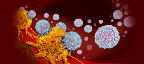
Cell Viability Assays: An Overview
Cell viability assays are essential tools in the fields of biology and medicine, offering researchers a reliable means to assess the health and viability of cells under various conditions. These assays play a crucial role in drug development, toxicology studies, and cancer research, enabling scientists to evaluate the effects of treatments and environmental factors on cell survival.
Understanding Cell Viability
Cell viability refers to the ability of cells to maintain metabolic functions and reproduce. Assessing cell viability is fundamental in many experimental setups, as it provides insights into cellular responses to drugs, nutrients, and stressors. Evaluating whether cells are alive or dead can inform researchers about the effectiveness of therapeutic agents and the cytotoxic potential of compounds.
Common Methods for Cell Viability Assessment
Various methods are employed in cell viability assays, each with its unique principles and applications. Here are some of the most widely used techniques:
Trypan Blue Exclusion: This traditional method involves staining cells with Trypan Blue, a dye that penetrates only dead cells. Viable cells remain unstained, allowing for easy differentiation. Researchers count the blue (dead) and clear (live) cells under a microscope.
Mitochondrial Activity Assays: These assays measure cellular respiration as an indicator of viability. Compounds such as MTT, XTT, or Resazurin are commonly used, wherein viable cells convert these dyes into colored products, indicating metabolic activity.
ATP Measurement: This method quantifies ATP levels in cells, as ATP is an essential energy molecule indicative of cell viability. Assays such as the ATP luminescence assay provide a sensitive measurement, often using bioluminescence to detect the emitted light.
Flow Cytometry: This advanced technique allows for the simultaneous analysis of multiple parameters in thousands of cells per second. Flow cytometry can differentiate live and dead cells by using fluorescent dyes and provides detailed information on cell populations.
Colorimetric and Fluorometric Assays: Colors or fluorescence intensity are measured following the addition of specific reagents to the cells. The intensity of the signal correlates with the number of viable cells, providing a quantitative assessment.
Applications of Cell Viability Assays
Cell viability assays are versatile and have numerous applications across various research domains:
Drug Development: In pharmacology, researchers use these assays to evaluate the cytotoxic effects of new drugs. Determining the effective concentration range and understanding dose-response relationships are critical for therapeutic development.
Toxicology: Assessing the impact of environmental toxins or chemicals on cell viability helps determine safety levels and mechanisms of toxicity.
Cancer Research: Understanding tumor cell response to treatment regimens is pivotal in developing effective cancer therapies. Cell viability assays help identify sensitive and resistant cancer cell lines, thereby guiding treatment strategies.
Stem Cell Studies: Evaluating the viability of stem cells is essential for regenerative medicine and developmental biology. These assays help confirm the health of stem cell populations before differentiation or transplantation.
Conclusion
Cell viability assays are invaluable tools in research, providing insights into cellular health and responses to various treatments. With advancements in technology, these assays continue to evolve, offering enhanced sensitivity and the ability to measure multiple parameters simultaneously. Understanding cell viability not only furthers our grasp of basic biological processes but also propels the development of new therapeutic strategies in medicine. Through these assays, researchers are equipped to unravel the complex interplay of cell survival, signaling, and the effects of external compounds, paving the way for innovations in health and disease management.
- Like
- Reply
-
Share
Reply
About Us · User Accounts and Benefits · Privacy Policy · Management Center · FAQs
© 2026 MolecularCloud


